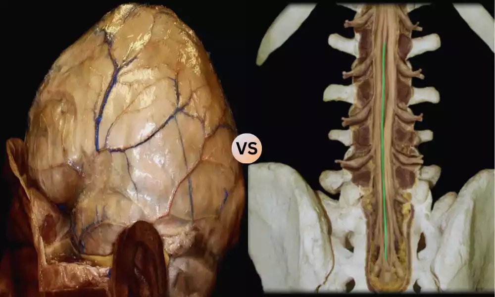
Cranial Dura and Spinal Dura, both components of the dura mater, are integral parts of the central nervous system’s protective covering. The cranial dura, encompassing the brain, features a double-layered structure with dural venous sinuses, serving as a crucial protector and structural support for the brain.
The spinal dura, continuous with its cranial counterpart but single-layered, envelops the spinal cord within the vertebral canal. It provides protection while allowing some degree of movement and flexibility. Understanding the distinctions between these two structures is essential in various clinical contexts, as each plays a unique role in safeguarding the central nervous system.
What is Cranial Dura?
The cranial dura, also known as the dura mater cranial or simply cranial dura, is a component of the meninges, which are the protective membranes that envelop the brain and spinal cord within the skull and vertebral canal. It is the innermost layer of the meninges surrounding the brain.
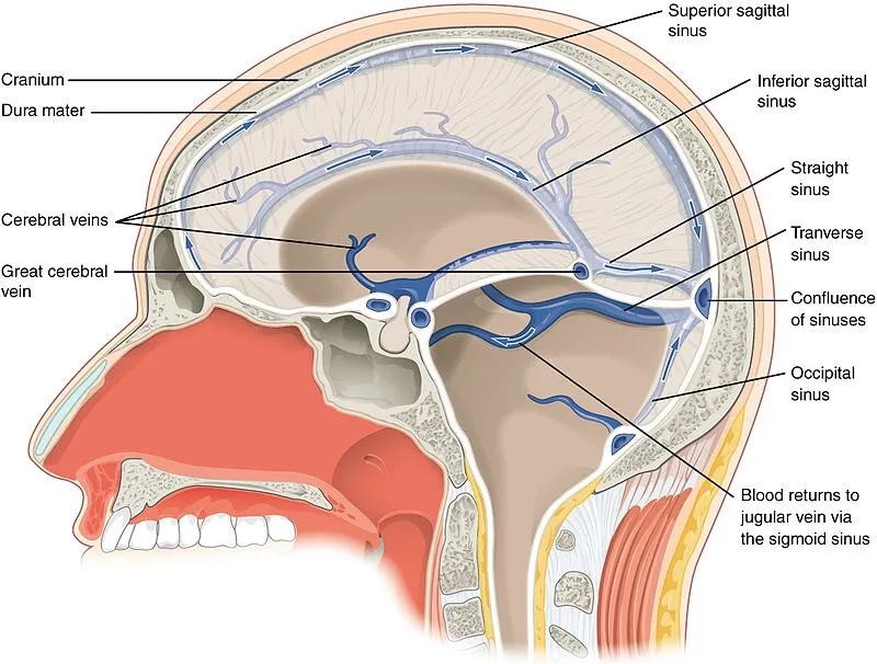
The cranial dura is a tough, fibrous membrane that consists of two layers the outer periosteal layer, which is attached to the inner surface of the skull bones, and the inner meningeal layer, which is closely adherent to the brain’s surface. Between these two layers are potential spaces known as dural venous sinuses, which play a role in the drainage of blood and cerebrospinal fluid.
The primary functions of the cranial dura are to protect the brain from external forces and provide structural support. It also contains various dural reflections and partitions that help compartmentalize and support different parts of the brain. Understanding the cranial dura is essential in neuroanatomy and neurosurgery due to its crucial role in safeguarding the brain.
What is Spinal Dura?
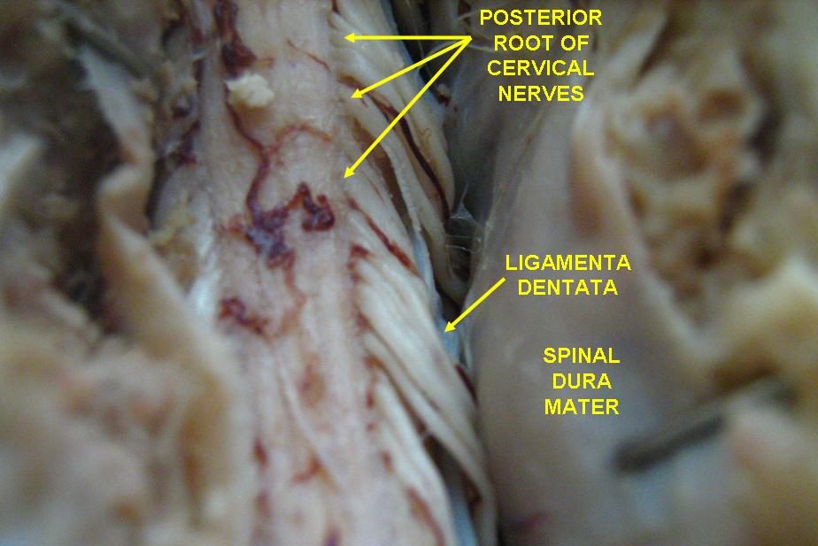
The spinal dura, also known as the dura mater spinalis or simply spinal dura, is a component of the meninges, which are the protective membranes that surround the spinal cord within the vertebral canal. It is continuous with the cranial dura, which envelops the brain, but it specifically encases the spinal cord.
The spinal dura is a tough, fibrous membrane that forms a single-layered sac around the spinal cord. It is situated within the vertebral canal, providing protection and cushioning for the spinal cord. Unlike the cranial dura, the spinal dura lacks the double-layered structure and potential spaces like the dural venous sinuses. Instead, it maintains a simpler, single-layered configuration.
One of the important functions of the spinal dura is to protect the spinal cord from mechanical injury and provide a degree of shock absorption. Additionally, the spinal dura plays a role in the containment and distribution of cerebrospinal fluid, which surrounds and bathes the spinal cord.
Understanding the anatomy and function of the spinal dura is essential in the fields of neurology, neurosurgery, and spinal medicine, as it is a critical component in the protection and support of the spinal cord.
Comparison Table of Cranial Dura and Spinal Dura
Here’s a comparison table highlighting the key differences between Cranial Dura and Spinal Dura:
| Characteristic | Cranial Dura | Spinal Dura |
|---|---|---|
| Location | Surrounds the brain within the cranial cavity | Surrounds the spinal cord within the vertebral canal |
| Layer Structure | Double-layered (periosteal and meningeal layers) | Single-layered |
| Potential Spaces | Contains dural venous sinuses | Lacks dural venous sinuses |
| Attachment to Bone | Attached to the inner surface of the skull bones | Not directly attached to the bone; continuous with cranial dura |
| Function | – Protection of the brain from external forces
– Structural support for the brain |
– Protection of the spinal cord from mechanical injury
– Shock absorption – Containment and distribution of cerebrospinal fluid |
| Clinical Significance | Relevant in cranial surgeries, dural venous sinus conditions, and intracranial pathology | Important in spinal surgeries, spinal cord injuries, and spinal disorders |
This table provides a concise overview of the primary differences between the cranial dura and spinal dura in terms of their location, structure, functions, and clinical significance.
Similarities of Cranial Dura and Spinal Dura
Cranial dura and spinal dura are both components of the meninges, which are the protective membranes that envelop various parts of the central nervous system. While they have distinct anatomical and functional differences, they also share some similarities:
-
- Composition: Both cranial dura and spinal dura are composed of a fibrous, dense connective tissue that provides durability and protection to the underlying neural structures.
- Meningeal Origin: Both the cranial dura and spinal dura originate from the same embryonic tissue, which means they share a common developmental origin.
- Connectivity: The cranial dura is continuous with the spinal dura at the foramen magnum, the large opening at the base of the skull where the spinal cord exits the cranial cavity and enters the vertebral canal. This continuity ensures a seamless transition from the cranial dura to the spinal dura.
- Cerebrospinal Fluid (CSF) Environment: Both the cranial dura and spinal dura play a role in maintaining the cerebrospinal fluid (CSF) environment around the central nervous system. While the cranial dura is more involved in the intracranial CSF dynamics, the spinal dura helps contain and distribute CSF along the spinal cord.
- Protection: Both membranes serve as protective barriers for the underlying neural tissues. The cranial dura protects the brain from external forces and provides structural support, while the spinal dura safeguards the spinal cord from mechanical injury.
- Clinical Considerations: In clinical practice, both cranial and spinal dura may be subject to various pathological conditions and surgical interventions. Understanding the similarities in their composition and functions is essential for neurosurgeons, neurologists, and other healthcare professionals involved in the treatment of neurological disorders.
While there are these similarities, it’s important to recognize that the cranial dura and spinal dura also have distinct characteristics and functions due to their different anatomical locations and roles within the central nervous system.
Clinical Significance
The clinical significance of understanding the differences between cranial dura and spinal dura, as well as their respective functions, extends to various medical and surgical contexts involving the central nervous system.
Here are some key clinical considerations:
- Neurosurgery: Neurosurgeons must have a deep understanding of the cranial dura and spinal dura when performing surgical procedures on the brain or spinal cord. Knowledge of their anatomy, attachment points, and potential spaces is crucial for minimizing complications during surgery and ensuring patient safety.
- Intracranial Pathology: Cranial dura can be affected by various pathological conditions, such as dural tears during cranial surgery or dural venous sinus thrombosis. Recognizing and addressing these issues is essential to prevent complications and ensure proper healing.
- Spinal Surgery: Spinal surgeons deal with the spinal dura during procedures such as laminectomies, spinal fusions, or the removal of spinal tumors. Understanding the spinal dura’s role in protecting the spinal cord and containing cerebrospinal fluid is critical in preventing damage during surgery.
- Spinal Cord Injuries: Spinal dura plays a role in spinal cord injuries. Understanding its structure and function helps healthcare professionals assess and manage spinal cord injuries effectively, whether they are traumatic or non-traumatic in nature.
- Spinal Disorders: Conditions like spinal stenosis, herniated discs, or spinal tumors can impact the spinal dura. Recognizing these conditions and their effects on the spinal dura is vital for accurate diagnosis and treatment planning.
- Cerebrospinal Fluid (CSF) Dynamics: The cranial dura, along with the spinal dura, contributes to the regulation of cerebrospinal fluid flow and pressure within the central nervous system. Disorders affecting CSF dynamics, such as hydrocephalus or CSF leaks, require an understanding of these membranes for proper management.
- Pain Management: The epidural space, which lies just outside the spinal dura, is a common site for pain management procedures such as epidural steroid injections. Understanding the anatomy and location of the spinal dura is essential for the safe and effective administration of these treatments.
- Diagnostic Imaging: Radiologists and clinicians use various imaging modalities, including MRI and CT scans, to visualize the cranial and spinal dura. Recognizing normal and abnormal appearances of these structures aids in diagnosing neurological conditions.
A thorough understanding of the cranial dura and spinal dura is critical in the fields of neurosurgery, neurology, radiology, and spinal medicine. It enables healthcare professionals to make accurate diagnoses, plan appropriate treatments, and minimize complications when dealing with conditions affecting the central nervous system.
Summary
Recognizing the distinctions between cranial dura and spinal dura is paramount in clinical practice. These differences, along with their shared anatomical and functional aspects, are vital for safe and effective management of central nervous system-related conditions, surgical procedures, and diagnostic evaluations. Understanding the clinical significance of these membranes enhances patient care and reduces the risk of complications.