
Hematoma and Hemangioma are two distinct medical terms referring to conditions related to blood but with different origins and characteristics. A hematoma arises when blood accumulates outside of blood vessels, usually due to trauma, leading to a localized collection that can cause swelling and discoloration. On the other hand, a hemangioma is a benign tumor composed of an abnormal proliferation of blood vessels. Often appearing as red or blue-tinted skin lesions, hemangiomas can grow rapidly, especially during infancy, and can be found anywhere on the body. While both conditions involve blood, their causes, presentations, and treatments vary significantly.
What is Hematoma?
A Hematoma is a localized collection of blood outside the blood vessels, usually caused by trauma, injury, or other disruptions to the integrity of blood vessels. It results in pooling of blood within tissues, leading to swelling and often manifesting as a bruise or discoloration on the skin. Hematomas can occur in various parts of the body, including the brain, muscles, and subcutaneous tissues, and they may vary in size from tiny spots to large masses. Depending on the location and size, hematomas can lead to pain, pressure on nearby structures, and other complications.

Causes of Hematoma
Hematomas form when blood leaks outside the vascular system, often as a result of damage to the blood vessels. Here are the primary causes of a hematoma:
- Trauma or Injury: The most common cause of hematomas.
- Blunt trauma: Such as a blow, fall, or bump.
- Penetrating trauma: Such as stab wounds or gunshot wounds.
- Medical Procedures:
- Surgery: Blood may pool at the surgical site.
- Blood draws or intravenous (IV) insertions: Especially if the needle punctures through the vein or if there’s significant movement during the procedure.
- Medications and Drugs: Certain medications can increase the risk.
- Anticoagulants (blood thinners): Like warfarin, heparin, or certain newer anticoagulant drugs.
- Antiplatelet drugs: Like aspirin and clopidogrel.
- Nonsteroidal anti-inflammatory drugs (NSAIDs): Such as ibuprofen or naproxen, when taken regularly or in high doses.
- Blood Disorders:
- Hemophilia: A genetic disorder where blood doesn’t clot normally.
- Platelet disorders: Conditions where the blood doesn’t clot effectively due to platelet dysfunction.
- Chronic Conditions:
- Liver disease: Liver problems can lead to a decreased production of clotting factors.
- Kidney disease: Can be associated with bleeding and bruising tendencies.
- Sports and Activities: Participation in contact sports or activities with high risk of injury can lead to frequent hematomas.
- Other Factors:
- Age: Elderly individuals tend to bruise more easily due to thinner skin and weaker blood vessels.
- Dietary deficiencies: Lack of certain nutrients, like vitamin K or vitamin C, can increase bleeding and bruising risk.
While hematomas are usually harmless and resolve on their own, some can lead to complications, especially if they occur in crucial areas like the brain or if they grow too large. Proper identification and management, when necessary, can help prevent any adverse outcomes.
Types of Hematoma
Here’s a concise breakdown of the main types of hematoma:
- Subdural Hematoma: Occurs between the brain’s dura mater and the arachnoid mater. Often caused by head trauma and can be life-threatening if it places pressure on the brain.
- Epidural Hematoma: Located between the skull and the dura mater of the brain. Typically results from a severe head injury, often associated with skull fractures.
- Intracerebral (or intraparenchymal) Hematoma: Found within the brain tissue itself. Can be due to trauma, stroke, or other conditions.
- Subarachnoid Hematoma: Occurs in the space between the arachnoid mater and the pia mater, often due to a traumatic event or aneurysm rupture.
- Intramuscular Hematoma: Located within muscle tissue, usually resulting from a direct blow or strain.
- Subcutaneous Hematoma: Found beneath the skin, often appearing as bruises.
- Perichondrial Hematoma: Forms on the ear’s cartilage, commonly referred to as “cauliflower ear.”
- Septal Hematoma: Occurs within the nasal septum, typically after trauma to the nose.
These are broad categories, and each type can have varying severity and implications depending on size, location, and cause.
Clinical Presentation of Hematoma
Here’s a concise breakdown of the clinical presentation of a hematoma:
- Swelling: Raised area where blood has pooled.
- Discoloration: Can range from red immediately after injury to purple, blue, green, yellow, or brown as it heals.
- Pain or Tenderness: At the site due to pressure from the pooled blood.
- Warmth: The area may feel warm to the touch.
- Limited Mobility: If located near a joint or in a muscle.
- Pressure Symptoms: For intracranial hematomas, symptoms like headaches, confusion, drowsiness, and even unconsciousness.
- Firmness: Over time, the pooled blood can cause the area to feel firm or rubbery.
The presentation can vary depending on the hematoma’s size, type, and location. Some internal hematomas might not have visible or palpable signs and might only be detected through symptoms or imaging.
Diagnosis of Hematoma
The diagnosis of a hematoma is primarily based on clinical assessment, but several diagnostic tools can be utilized, especially for internal hematomas. Here’s a concise breakdown of how hematomas are diagnosed:
- Physical Examination:
- Inspection of swelling, discoloration, and palpation for tenderness or firmness.
- Neurological examination, particularly for suspected intracranial hematomas, to check for altered consciousness, pupil response, and other neurological deficits.
- Medical History:
- Gathering information on the incident leading to the injury, previous similar episodes, current medications (especially anticoagulants), and any underlying health conditions.
- Imaging Studies:
- X-ray: Useful for ruling out fractures, which can accompany hematomas.
- Ultrasound: Effective for visualizing soft tissue hematomas, such as those in muscles. It can differentiate between old and fresh hematomas and identify any associated injuries.
- Computed Tomography (CT) Scan: Particularly useful for diagnosing intracranial hematomas. Provides a clear view of the hematoma’s location and size.
- Magnetic Resonance Imaging (MRI): Offers a detailed view of tissues, making it useful for brain hematomas and those in musculoskeletal regions.
- Blood Tests:
- To check platelet count, clotting factors, and other parameters if an underlying bleeding disorder is suspected.
- Aspiration:
- In cases where the diagnosis is uncertain, a needle may be used to draw out some of the fluid for analysis.
Once a hematoma is diagnosed, the healthcare provider will determine the severity and decide on the appropriate course of treatment.
Treatment for Hematoma
Treatment for a hematoma depends on its size, type, location, and any associated complications. Here’s a concise breakdown of the common treatments for hematomas:
- Conservative Management:
- Rest: Limiting activity can help prevent the hematoma from growing.
- Ice: Applying cold compresses can reduce swelling and alleviate pain.
- Compression: Using elastic bandages can help minimize swelling.
- Elevation: Elevating the affected area above the level of the heart helps reduce swelling.
- Medication:
- Pain Relievers: Over-the-counter pain medications like acetaminophen or nonsteroidal anti-inflammatory drugs (NSAIDs) such as ibuprofen can help manage pain and inflammation.
- Review of Current Medications: If a patient is on blood thinners and recurrent hematomas are an issue, the doctor might reconsider the dose or the medication itself.
- Aspiration:
- For larger hematomas that are causing discomfort or pressure, a doctor might use a needle to draw out the pooled blood. This procedure provides immediate relief.
- Surgical Drainage:
- Used for larger hematomas or those causing complications. Particularly common for certain types of intracranial hematomas that cause increased pressure on the brain.
- Insertion of a drainage tube might be necessary in some cases.
- Surgical Repair:
- In cases where a blood vessel is actively bleeding or has been damaged significantly, surgical intervention might be necessary to repair it.
- Observation:
- Some small hematomas, especially subcutaneous ones, might not need active treatment and can resolve on their own over time.
- Physical Therapy:
- For hematomas that affect joints or muscles, physical therapy might be recommended to restore full function.
It’s crucial for patients to consult with a healthcare professional for an accurate diagnosis and appropriate treatment. If a hematoma is suspected, especially in critical areas like the brain, timely medical attention is imperative.
Complications of Hematoma
Here’s a concise breakdown of the potential complications of a hematoma:
- Infection: Especially if the skin is broken or if the hematoma is due to a penetrating injury.
- Compression: Hematomas can press on nearby structures, nerves, or blood vessels, causing pain or functional impairment.
- Calcification: Over time, some hematomas may calcify, leading to a hard lump.
- Prolonged Inflammation: Persistent hematomas can lead to chronic inflammation in the area.
- Cosmetic Concerns: Large or visible hematomas can result in scarring or discoloration.
- Abscess Formation: Infected hematomas can turn into abscesses.
- Neurological Complications: Intracranial hematomas can lead to increased intracranial pressure, brain damage, or even death if not treated promptly.
- Compartment Syndrome: Hematomas within muscle compartments can raise the pressure, reducing blood flow and potentially damaging muscles and nerves.
It’s essential to monitor hematomas for any signs of these complications and seek medical intervention when needed.
What is Hemangioma?
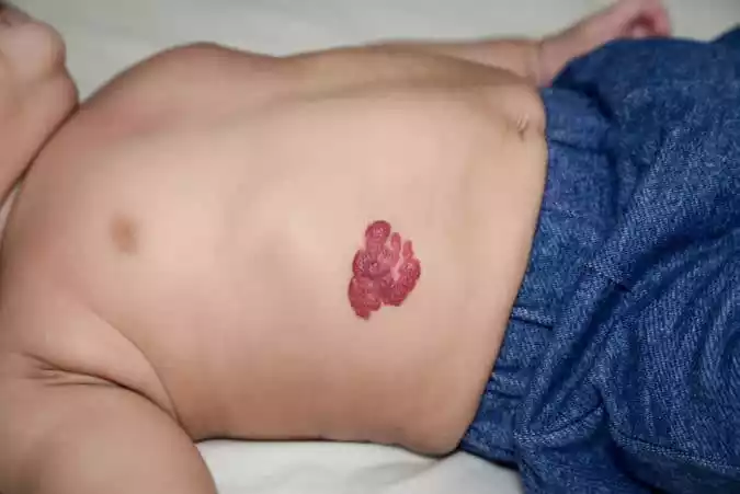
A Hemangioma is a benign (non-cancerous) tumor made up of an abnormal proliferation of blood vessels. It can appear at birth or develop in the first or second week of life. A hemangioma can occur anywhere on the body, but most commonly appears on the face, scalp, chest, or back. While the exact cause of hemangiomas is not fully understood, they tend to grow rapidly during the first year of life and then undergo a phase of regression. Most hemangiomas will have disappeared or faded considerably by the time a child reaches the age of 10.
Causes of Hemangioma
The exact cause of hemangiomas is not fully understood, but they are believed to result from a combination of genetic and environmental factors. Here are some insights into the potential causes and associated factors:
- Vascular Anomalies: Hemangiomas are a type of vascular anomaly, which means they are related to irregularities in the formation or development of blood vessels.
- Placental Hypothesis: Some research suggests that hemangiomas might originate from placental tissue. Certain cells found in hemangiomas are similar to those found in the placenta.
- Hormonal Influence: The rapid growth of some hemangiomas in the first year of life has led researchers to hypothesize that hormones might play a role, though this is not confirmed.
- Genetic Factors: While the majority of hemangiomas occur sporadically, there is evidence that they can run in families, suggesting a possible genetic component.
- Environmental Factors: Some studies have suggested that certain conditions during pregnancy, such as placental complications or pre-eclampsia, might increase the risk of a baby developing a hemangioma.
- Other Associated Factors: Premature birth and low birth weight have been linked to an increased risk of infantile hemangiomas.
It’s essential to understand that while these are potential causes and associated factors, the definitive cause of hemangiomas remains unclear. They are not caused by anything a mother did or did not do during pregnancy, and they are not a result of an injury or trauma.
Types of Hemangioma
Here’s a concise breakdown of the main types of hemangiomas:
- Infantile Hemangioma:
- Most common type, appearing soon after birth or within the first month of life.
- Often undergo a rapid growth phase followed by a spontaneous involution phase, where it gradually fades.
- Congenital Hemangioma:
- Present at birth.
- Two main types: Rapidly Involuting Congenital Hemangioma (RICH) which disappears within the first year of life, and Non-Involuting Congenital Hemangioma (NICH) which remains stable.
- Capillary Hemangioma (Strawberry Hemangioma):
- The most common type of infantile hemangioma.
- Appears as a raised, red, strawberry-like lesion.
- Cavernous Hemangioma:
- Composed of larger, dilated blood vessels.
- Deeper in the skin and may appear as a bluish mass.
- Compound Hemangioma:
- Combines features of both capillary and cavernous hemangiomas.
- Internal Hemangioma:
- Develops inside the body, often in the liver, brain, or other internal organs.
- Might not be detected unless they cause symptoms.
Each type can vary in size, location, and appearance. While hemangiomas are generally benign, their growth can sometimes cause complications, depending on their location.
Clinical Presentation of Hemangioma
Here’s a concise breakdown of the clinical presentation of a hemangioma:
- Coloration:
- Red to bluish tint, depending on depth in the skin.
- Texture:
- Smooth or lumpy on the skin’s surface.
- Size and Growth:
- Can be tiny or several inches across.
- May exhibit rapid growth, especially during the first year of life.
- Location:
- Commonly on the face, scalp, chest, or back, but can occur anywhere on the body or even internally.
- Symptoms (for internal hemangiomas):
- May be asymptomatic or cause symptoms related to the affected organ.
- Warmth:
- The area might feel warm due to increased blood flow.
- Pulsation:
- Rarely, a large hemangioma might have a noticeable pulse.
- Ulceration:
- In some cases, the surface of the hemangioma can break down, leading to ulceration.
- Pain or Discomfort:
- Especially if the hemangioma is in a location where it’s subjected to frequent friction or if it becomes ulcerated.
The presentation can vary widely depending on the type, size, and location of the hemangioma.
Diagnosis of Hemangioma
The diagnosis of a hemangioma often starts with a physical examination and a review of the patient’s medical history. Here’s a concise breakdown of the diagnostic process for hemangiomas:
- Physical Examination:
- Inspection of the lesion’s size, color, and texture.
- Palpation to assess depth and consistency.
- Medical History:
- Information about when the hemangioma first appeared and its growth pattern.
- Imaging Studies:
- Ultrasound: Helps distinguish hemangiomas from other vascular lesions and can provide details about depth and blood flow.
- Magnetic Resonance Imaging (MRI): Useful for larger, deep, or internal hemangiomas. Provides detailed images of the lesion and its relationship to nearby structures.
- Computed Tomography (CT) Scan: Sometimes used for internal hemangiomas, especially in the liver.
- Biopsy:
- Rarely done, as most hemangiomas can be diagnosed based on their appearance and imaging studies. However, if there’s uncertainty about the diagnosis or concerns about other conditions, a small sample might be taken for histopathological examination.
- Other Tests for Internal Hemangiomas:
- Depending on the location, specific tests like liver function tests (for hepatic hemangiomas) may be conducted.
It’s important to differentiate hemangiomas from other vascular lesions and tumors. While hemangiomas are benign, accurate diagnosis ensures appropriate management and monitoring.
Treatment for Hemangioma
The treatment for hemangiomas depends on the size, location, type, and any associated symptoms or complications. Here’s a concise breakdown of the common treatments for hemangiomas:
- Observation:
- Many hemangiomas don’t require treatment and will fade on their own, especially infantile hemangiomas which often undergo spontaneous involution.
- Topical Treatments:
- Beta-blockers: Topical timolol gel can be used for superficial hemangiomas, reducing their size and redness.
- Steroids: Topical or injected steroids can help reduce the size of a hemangioma.
- Oral Medications:
- Propranolol: An oral beta-blocker, is the primary treatment for problematic infantile hemangiomas. It can reduce the size, color, and appearance.
- Steroids: Oral corticosteroids may be used for large or problematic hemangiomas, especially if they are interfering with organ function or causing other complications.
- Other medications: Such as vincristine or interferon alfa, are sometimes used for hemangiomas that don’t respond to other treatments.
- Laser Therapy:
- Pulsed dye laser can be effective in treating superficial hemangiomas by reducing their redness and stopping their growth.
- Surgery:
- Surgical removal is considered for hemangiomas that cause functional problems, are very large, or for cosmetic reasons. It’s also an option for hemangiomas that don’t respond to other treatments.
- Sclerotherapy:
- Involves injecting a solution into the hemangioma to shrink it. It’s often used for larger, cavernous hemangiomas.
- Embolization:
- A procedure where the blood vessels feeding the hemangioma are blocked, causing it to shrink. Often used for large or internal hemangiomas, especially those in the liver.
It’s essential for patients to consult with a healthcare professional to determine the best course of action based on the individual characteristics and complications of the hemangioma.
Complications of Hemangioma
Here’s a concise breakdown of potential complications associated with hemangiomas:
- Ulceration:
- The surface of the hemangioma can break down, leading to open sores that can be painful and prone to infection.
- Bleeding:
- If injured or ulcerated, a hemangioma can bleed.
- Disfigurement:
- Depending on its size and location, a hemangioma can lead to cosmetic concerns.
- Vision or Breathing Issues:
- Hemangiomas near the eyes, nose, or throat can interfere with vision or breathing.
- Internal Complications:
- Hemangiomas in the liver, for instance, can cause complications such as abdominal pain or bleeding.
- Psychological Impact:
- Large or visible hemangiomas can affect a person’s self-esteem and social interactions.
- Heart Issues:
- Rarely, a large hemangioma can put stress on the heart due to increased blood flow.
- Compression:
- Hemangiomas growing in certain areas might compress adjacent structures, nerves, or organs.
It’s essential to monitor hemangiomas, especially those in critical areas or of significant size, to ensure that potential complications are addressed promptly.
Comparison table of Hematoma and Hemangioma
Here’s a comparison table highlighting the differences between hematoma and hemangioma:
| Aspect | Hematoma | Hemangioma |
|---|---|---|
| Definition | A localized collection of blood outside of blood vessels. | A benign tumor made up of an abnormal proliferation of blood vessels. |
| Cause | Usually due to trauma, injury, surgery, or a medical condition causing bleeding. | Unknown; possibly due to a combination of genetic and environmental factors. |
| Appearance | Bruise-like discoloration; can be swollen and raised. | Red to bluish tumor; varies in size. Can be superficial or deep. |
| Location | Can occur anywhere in the body where bleeding occurs, including skin, muscles, and internal organs. | Commonly on the face, scalp, chest, or back but can occur anywhere, including internally. |
| Onset | Rapid onset after trauma or injury. | Can appear at birth or within the first month of life. |
| Duration | Typically temporary; resolves as the body reabsorbs the blood. | Can grow during the first year of life, then may fade over years. Some remain unchanged. |
| Treatment | RICE (rest, ice, compression, elevation), pain relievers, aspiration, or surgical drainage in severe cases. | Observation, oral medications, laser therapy, surgery, or other treatments depending on size, location, and complications. |
| Complications | Infection, compression, prolonged inflammation, calcification. | Ulceration, bleeding, cosmetic concerns, vision or breathing issues, internal complications, psychological impact. |
This table provides a general comparison, and individual cases may vary. Always consult with a healthcare professional for accurate diagnosis and treatment.
Preventive Measures of Hematoma and Hemangioma
Preventing hematomas and hemangiomas differs due to the distinct nature of their causes. Here are preventive measures for both:
Hematoma:
- Protective Gear: Wear appropriate protective gear during sports or activities with a risk of physical injury.
- Avoid Falls: For the elderly or those with balance issues, ensure that the living environment is safe from trip hazards. Using aids like handrails can help.
- Medication Caution: If on anticoagulant or antiplatelet medications, be cautious of activities that might lead to bruising or bleeding.
- Monitor Medical Procedures: After surgeries or medical procedures, follow post-operative care instructions to reduce bleeding risks.
- Treat Medical Conditions: Conditions that affect blood clotting or platelet function should be managed effectively.
- Avoid Excessive Alcohol: Excessive alcohol can interfere with blood clotting.
Hemangioma:
- Genetic Counseling: If there’s a family history of hemangiomas, genetic counseling might offer insights into potential risks for future offspring.
- Optimal Pregnancy Health: While the direct cause of hemangiomas remains unknown, maintaining optimal health during pregnancy can reduce general risks. This includes avoiding tobacco, limiting alcohol, and following a doctor’s advice.
- Monitor Newborns: Keeping an eye on any new skin growths in infants allows for early detection and potential intervention if needed.
- Regular Check-ups: Especially for internal hemangiomas, regular health check-ups can detect any issues early.
While these measures can reduce risks, they might not completely prevent the development of hematomas or hemangiomas. Always consult a healthcare professional for guidance tailored to individual circumstances.
Conclusion
Hematoma and hemangioma, though both related to blood vessels, represent distinct medical conditions. A hematoma is a localized collection of blood outside of blood vessels, usually due to trauma or bleeding disorders. In contrast, a hemangioma is a benign tumor arising from an abnormal proliferation of blood vessels, commonly found on the skin or internally.
While hematomas result from injuries or underlying medical conditions and are generally temporary, the exact cause of hemangiomas remains uncertain, and they can persist for longer durations. Both conditions require different approaches to management, but with early detection and appropriate care, complications can be minimized.