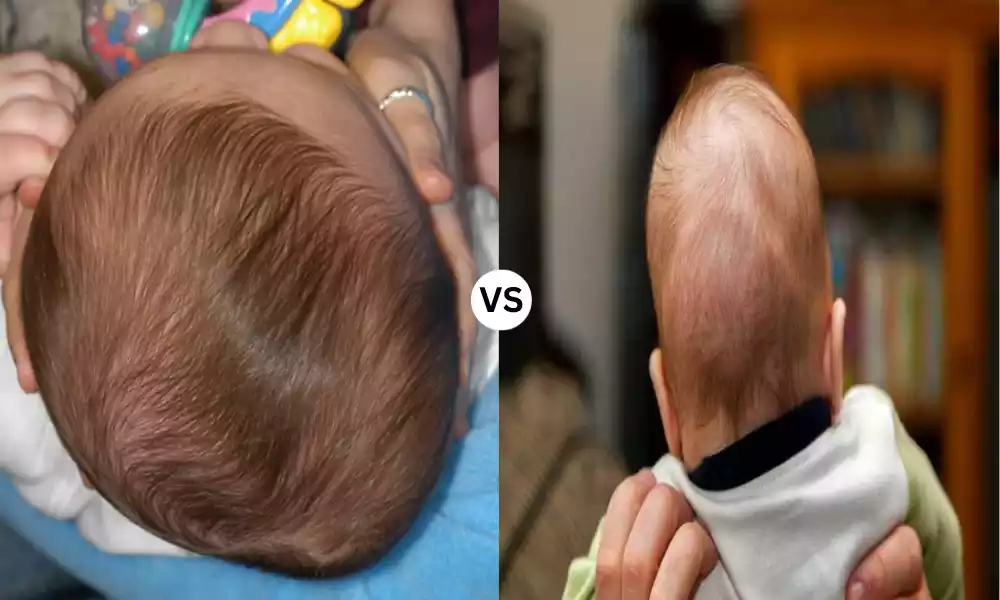
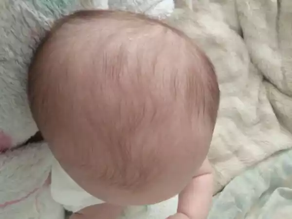
Plagiocephaly and Craniosynostosis are conditions that affect the shape and structure of an infant’s skull. Plagiocephaly, often referred to as “flat head syndrome,” is characterized by a flattened spot on the back or side of an infant’s head, typically resulting from prolonged pressure in one position. It is often a cosmetic concern and can usually be addressed with non-surgical interventions. On the other hand, craniosynostosis is a more serious condition where one or more of the fibrous joints (sutures) between the bones of a baby’s skull close prematurely.
This can restrict the brain’s growth, leading to potential developmental issues and requiring surgical intervention. Both conditions underscore the importance of early detection and the need for parental awareness and education.
What is Plagiocephaly?
Plagiocephaly, commonly known as “flat head syndrome,” refers to the asymmetrical flattening of one side of an infant’s head. This condition can develop due to the baby’s position in the womb or from regularly resting in a certain position during infancy, especially in the early months when the skull is soft and malleable.
There are two main types of plagiocephaly:
- Positional (or Deformational) Plagiocephaly: This is the most common form. It occurs when an infant’s head becomes flattened due to external pressures. This might arise from consistently lying in one position, such as on their back. As a result, there is a flat spot on the back or side of the head, and there might be some slight asymmetry in the face, like a misalignment of the ears.
- Synostotic Plagiocephaly: This form is less common and occurs when the lambdoid suture on the back of the head fuses prematurely. It is a type of craniosynostosis, which means that it’s related to the early fusion of the sutures in the skull. It is more serious than positional plagiocephaly and requires surgical intervention.
Positional plagiocephaly is typically a cosmetic concern and does not affect brain development. With early intervention, such as repositioning the baby’s head regularly and allowing for “tummy time” when awake, the shape of the head can often be normalized. In more pronounced cases, physical therapy or helmet therapy might be recommended.
It’s important for caregivers to recognize the signs of plagiocephaly early on and to consult with pediatricians or specialists to determine the best course of action.
Causes of Plagiocephaly
The development of plagiocephaly, particularly positional or deformational plagiocephaly, is typically the result of external pressures on the soft and malleable skull of an infant. Here are the primary causes:
- Prolonged Pressure on One Side of the Head: Infants who lie in one position for extended periods, especially on their backs, may develop a flattened spot on that side of the head. The “Back to Sleep” campaign, which recommends placing babies on their backs to sleep to reduce the risk of Sudden Infant Death Syndrome (SIDS), has decreased the incidence of SIDS but has been associated with an increase in positional plagiocephaly.
- Premature Birth: Premature babies often have softer skull bones than full-term infants. Additionally, they might spend more time lying down without being moved or repositioned, especially if they are in neonatal intensive care units. This makes them more susceptible to developing plagiocephaly.
- Restrictive Intrauterine Position: During pregnancy, especially in cases of multiple births (twins, triplets, etc.), the womb can become cramped, causing an infant’s head to be pressed against the mother’s pelvis or the sibling’s body, leading to a flat spot.
- Neck Muscle Tightness (Torticollis): Some babies may have a condition called congenital muscular torticollis, where one of the neck muscles is tighter than the other. This condition can cause the baby to tilt or turn their head in one direction more frequently, leading to consistent pressure on one side of the skull.
- Use of Car Seats, Swings, and Bouncers: Extended periods in car seats, swings, and bouncers, where the baby’s head rests against a flat surface, can also contribute to the development of plagiocephaly.
- Position During Feeding: Consistently feeding the baby from the same side or position can lead to a preference for turning the head one way, contributing to a flat spot on that side.
While the causes listed above pertain primarily to positional or deformational plagiocephaly, synostotic plagiocephaly (a subset of craniosynostosis) results from the premature fusion of the lambdoid suture in the skull. The causes for this are different and often involve genetic or developmental factors.
Treatment Options
The treatment for plagiocephaly often depends on the severity of the condition and the age at which it’s identified. Here are the primary treatment options:
- Repositioning (Counter-Positioning):
- This is the first line of treatment, especially for younger infants.
- The goal is to reduce pressure on the flattened area by regularly changing the baby’s position.
- Encouraging “tummy time” when the baby is awake and supervised can help.
- Altering the baby’s position during sleep (while still ensuring they are on their back) can help. This might include changing the direction the baby faces in the crib or alternating the arm they lie on during feedings.
- Physical Therapy:
- For babies with neck muscle tightness or torticollis, physical therapy can be beneficial.
- A physical therapist can guide exercises to stretch and strengthen the neck muscles, which can help the baby turn their head more freely.
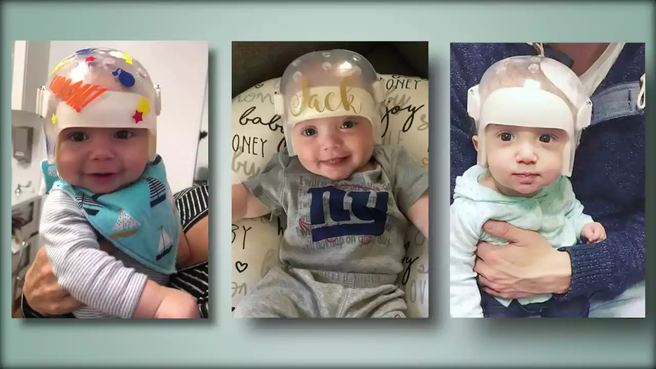
- Helmet or Cranial Orthosis Therapy:
- Used for moderate to severe cases of plagiocephaly.
- Custom-made helmets or headbands can be designed to fit over the baby’s skull. These devices apply gentle pressure to the bulging areas of the head and relieve pressure from the flattened areas, promoting more symmetrical growth.
- Typically, helmet therapy is most effective when started between the ages of 4 to 6 months and can last several months. It’s less effective after age 1 when the skull becomes more rigid.
- Cranial Surgery (Rare):
- While surgery is not typically required for positional plagiocephaly, severe cases of synostotic plagiocephaly (related to craniosynostosis) may require surgical intervention.
- The procedure involves reshaping the affected parts of the skull and is usually performed in the first year of life.
- Observation:
- Mild cases of plagiocephaly may self-correct as the baby grows, especially if the child becomes more active and spends less time lying down.
- Regular pediatric appointments can help monitor the condition to see if it’s improving on its own or if intervention is necessary.
- Education and Prevention:
- Educating parents about the importance of varied head positioning, tummy time, and avoiding extended time in car seats or bouncers can prevent or reduce the severity of plagiocephaly.
Remember, each child is unique, and treatment decisions should be made in consultation with pediatricians or specialists who can provide guidance tailored to the individual needs and circumstances of the child.
What is Craniosynostosis?
Craniosynostosis is a birth defect characterized by the premature fusion of one or more of the fibrous joints (sutures) between the bones in an infant’s skull. This abnormal fusion restricts the growth of the skull, potentially leading to increased intracranial pressure, developmental complications, and aesthetic concerns.
Key features of craniosynostosis include:
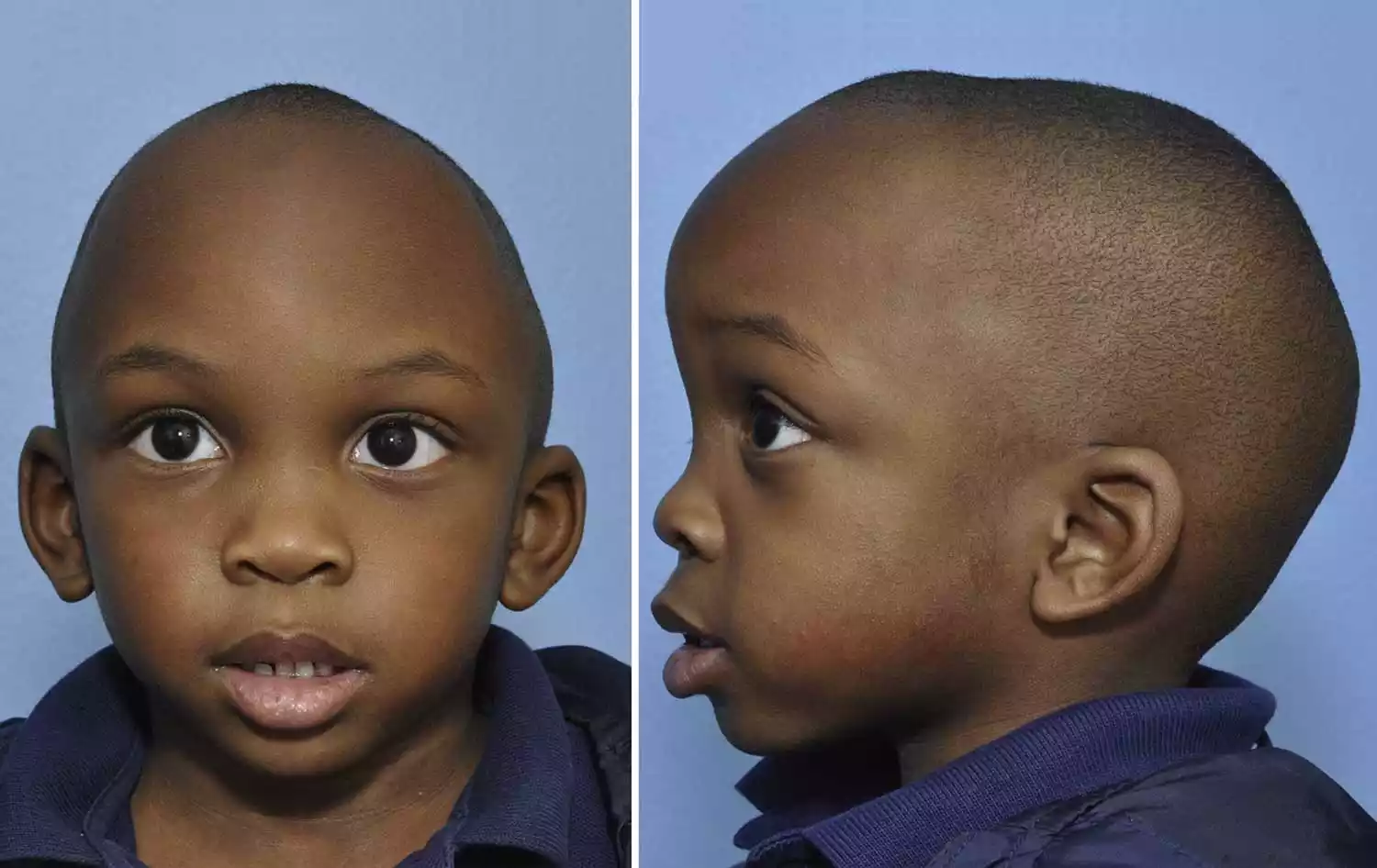
- Misshapen Skull: Depending on which suture(s) fuse early, the skull can have various abnormal shapes. For instance, fusion of the sagittal suture might result in a long, narrow head, while fusion of a coronal suture could cause a flattened or protruding forehead.
- Ridges or Absence of Soft Spots: The affected sutures may feel like raised, hard ridges. Conversely, the soft spots (fontanelles) might be absent or smaller than usual.
- Increased Intracranial Pressure: As the brain continues to grow, restricted skull space can lead to increased pressure within the skull. Symptoms may include irritability, bulging eyes, prominent veins on the scalp, and developmental delays.
- Related Genetic Syndromes: In some cases, craniosynostosis is part of a syndrome, such as Crouzon, Apert, or Pfeiffer syndrome. These syndromes often have other associated features, like facial deformities or limb abnormalities.
- Differing Types Based on Affected Sutures: Types include sagittal (scaphocephaly), coronal (anterior plagiocephaly or brachycephaly), lambdoid (posterior plagiocephaly), and metopic (trigonocephaly) synostosis.
- Treatment Requirement: Surgical intervention is typically required to allow for normal brain growth, relieve increased intracranial pressure, and improve cosmetic appearance.
Early diagnosis and intervention are paramount, as they offer the best chance for optimal outcomes, both functionally and aesthetically.
Causes of Craniosynostosis
Craniosynostosis is the premature fusion of one or more of the sutures (fibrous joints) in an infant’s skull. When these sutures close too early, it can restrict the growth of the skull in the affected areas and may force the skull to grow in other directions, potentially leading to an abnormal head shape and, in severe cases, increased intracranial pressure.
The causes of craniosynostosis can be multifaceted:
- Genetic Causes: Some cases of craniosynostosis are associated with genetic mutations or syndromes. Common syndromes linked to craniosynostosis include:
- Apert syndrome
- Crouzon syndrome
- Pfeiffer syndrome
- Carpenter syndrome
- Saethre-Chotzen syndrome The genes commonly associated with these syndromes play roles in bone development and growth.
- Environmental Factors: Some studies suggest that certain environmental factors during pregnancy might increase the risk of craniosynostosis. These factors include maternal smoking, certain infections during pregnancy, and drug use (especially certain anti-seizure medications).
- Metabolic Disturbances: Conditions that influence calcium and phosphate metabolism, such as hyperthyroidism and rickets, have been linked to craniosynostosis.
- Fetal Head Constraint: This refers to cases where the fetus’s head is restricted and unable to grow normally, such as in cases where there are multiple pregnancies (like twins or triplets) or where there’s oligohydramnios (low amniotic fluid). When the baby’s head is constricted or pressed against the mother’s pelvis, it can lead to positional molding that may be mistaken for craniosynostosis. However, true craniosynostosis is the premature fusion of the sutures.
- Idiopathic: Many cases of craniosynostosis have no known cause and are termed as idiopathic.
Every abnormal head shape in an infant is due to craniosynostosis. Positional plagiocephaly, which results from external pressures on the soft infant skull (like lying in one position for extended periods), can also lead to an abnormal head shape but does not involve suture fusion.
Treatment Options
The treatment of craniosynostosis primarily focuses on relieving pressure on the brain, ensuring that the brain has room to grow normally, and improving the cosmetic appearance of the head. The specific treatment approach varies depending on the suture(s) involved, the age of the child, the presence of associated syndromes, and the severity of the condition. Here are the primary treatment options:
- Surgery: Surgery is the most common and effective treatment for craniosynostosis.
- Traditional (Open) Surgery: This involves making an incision on the skull to open up the fused suture. Bone segments might be reshaped, repositioned, or even removed and then reattached using plates and screws that are often resorbable. This procedure is typically done in infants older than six months.
- Endoscopic Surgery: This is a less invasive procedure where small incisions are made, and an endoscope is used to release the fused suture. This procedure is usually done in younger infants, typically before 3-4 months of age. After the surgery, the infant often wears a custom-fitted helmet for several months to guide the reshaping of the skull.
- Spring-Assisted Surgery: In this method, springs are inserted to help expand the sutures and allow for more natural growth. Like endoscopic surgery, a molding helmet might be used postoperatively.
- Molding Helmets: These are specially designed helmets that apply gentle, consistent pressure to mold the baby’s skull into a more typical shape. They’re often used after endoscopic surgery to guide the skull’s growth. However, they are not effective for treating craniosynostosis by themselves but can be useful for addressing positional plagiocephaly, which is often confused with craniosynostosis.
- Observation: In very mild cases, where the fusion doesn’t threaten brain growth or development and the cosmetic appearance is not significantly affected, the physician might choose to monitor the child without immediate intervention. However, this approach is less common.
- Physical Therapy: In cases where positional preference or torticollis (a condition where the neck muscles cause the head to tilt in one direction) contributes to the head shape, physical therapy might be recommended to improve neck muscle balance and guide the child in varying head positions.
- Genetic Counseling: If craniosynostosis is associated with a genetic syndrome, genetic counseling might be beneficial for the family. This can help parents understand the risk of recurrence in future pregnancies and provide information on the specific syndrome.
It’s crucial to have a multidisciplinary team involved in the care of a child with craniosynostosis. This team often includes a pediatric neurosurgeon, a craniofacial/plastic surgeon, a pediatrician, an ophthalmologist (for vision concerns), a geneticist (especially if a syndrome is suspected), and other specialists as needed.
Comparison Table of Plagiocephaly and Craniosynostosis
Here’s a comparison table that highlights the primary differences between plagiocephaly and craniosynostosis:
| Feature | Plagiocephaly (Positional) | Craniosynostosis |
|---|---|---|
| Cause | External pressure on the soft infant skull, often due to baby lying in one position for extended periods. | Premature fusion of one or more cranial sutures. |
| Sutures Affected | No suture fusion. The soft spots (fontanelles) remain open and flexible. | One or more sutures fuse prematurely, leading to restricted skull growth in the affected area. |
| Head Shape | Flattening of one side of the head (typically the back or side). Can be anterior or posterior plagiocephaly. | Abnormal head shape varies based on the suture fused. Examples: scaphocephaly, trigonocephaly, brachycephaly etc. |
| Treatment | Molding helmets or headbands to help reshape the skull, and physical therapy if torticollis is present. | Surgery is the primary treatment, followed by molding helmets in some cases. |
| Age of Diagnosis | Often noticed a few weeks to months after birth. | Can be noticed at birth or soon after, especially if significant. |
| Associated Symptoms | Generally, no symptoms other than the flattened head shape. No increased intracranial pressure or other health concerns. | Increased intracranial pressure, prominent veins, developmental delays, etc., in severe cases. |
| Genetics | Typically not linked to genetic conditions. | Can be associated with genetic syndromes such as Apert, Crouzon, Pfeiffer, etc. |
It’s essential to get a correct diagnosis if you suspect either condition, as the treatment and prognosis can be very different.
Importance of Early Detection
Early detection of craniosynostosis, plagiocephaly, and other cranial deformities is crucial for several reasons:
- Optimal Outcomes with Minimally Invasive Procedures: Early detection, especially in craniosynostosis, may allow for less invasive surgical techniques, such as endoscopic surgery. These less invasive procedures often have shorter operative times, reduced blood loss, and faster recovery compared to traditional open surgeries. They need to be performed at a younger age, typically before 3-4 months.
- Improved Cosmetic Results: Early intervention can lead to better cosmetic outcomes by allowing the skull to grow and develop more normally. Whether through surgical correction or the use of molding helmets, the goal is to enable the child’s head to have a more typical appearance as they grow.
- Prevention of Potential Complications: In the case of craniosynostosis, untreated premature suture fusion can lead to increased intracranial pressure, which can have detrimental effects on the developing brain. Early detection and intervention can prevent or reduce these potential complications.
- Avoidance of Developmental Delays: Increased intracranial pressure from untreated craniosynostosis can lead to developmental delays. Addressing the issue early can ensure that the child’s brain has adequate space to grow, reducing the risk of such delays.
- Early Start on Physical Therapy: For plagiocephaly related to torticollis (tight neck muscles), early detection allows for the commencement of physical therapy to address the muscle tightness, which can, in turn, help in the resolution of the head shape abnormalities.
- Guided Skull Growth: Molding helmets, used either post-operatively for craniosynostosis or as a primary treatment for plagiocephaly, are most effective when started early, typically before the age of one year. This is because the infant’s skull is still very malleable, allowing for more effective reshaping.
- Reduction in Family Anxiety: Recognizing and addressing cranial deformities early can provide relief and clarity to parents and caregivers who might be anxious about their child’s appearance or potential health implications. Being proactive can also provide a clearer path forward in terms of treatment and expected outcomes.
- Prevention of Misdiagnosis or Delayed Diagnosis: Early detection ensures that conditions like craniosynostosis are not misdiagnosed as plagiocephaly or vice versa. The treatments are different, so it’s essential to have an accurate diagnosis from the outset.
In essence, early detection and intervention in craniosynostosis and plagiocephaly can lead to better functional, developmental, and cosmetic outcomes for the child. Regular pediatric check-ups and monitoring of the infant’s head shape and growth can aid in early detection.
Prevention and Care
While craniosynostosis often has genetic components or other underlying causes that make prevention challenging, plagiocephaly (positional) is more amenable to preventive strategies. Here are some measures for prevention and care related to plagiocephaly and general cranial care:
Plagiocephaly (Positional) Prevention:
- Tummy Time: Encourage “tummy time” while the infant is awake and under supervision. This not only helps strengthen neck muscles but also reduces the constant pressure on the back of the baby’s head when lying on their back.
- Alternate Head Positioning: When the baby is sleeping or resting on their back, try to alternate the direction the baby’s head turns. This prevents continuous pressure on one particular side of the head.
- Change Location of Toys and Stimuli: Rotate the position of toys, mobiles, or other visual stimuli around the crib or play area to encourage the baby to turn their head in different directions.
- Limit Time in Car Seats and Bouncy Seats: While it’s essential to use car seats for safety while driving, try to reduce the baby’s time in car seats, bouncers, or swings that confine them in one position.
- Hold Your Baby: Holding your baby upright over your shoulder or using baby carriers can help reduce the time the baby’s head is against a flat surface.
- Check for Torticollis: If you notice that your baby consistently tilts or turns their head to one side, they might have torticollis (tight neck muscles). Physical therapy can be effective in these cases.
Craniosynostosis Care:
- Regular Medical Check-ups: Regular visits to the pediatrician can help monitor the baby’s head growth and shape, ensuring early detection of any abnormalities.
- Genetic Counseling: If there’s a family history of craniosynostosis or if the child has a known associated syndrome, consider genetic counseling to understand the risks and implications better.
- Post-operative Care: If surgery is performed to correct craniosynostosis, it’s essential to follow post-operative care guidelines closely, including:
- Monitoring for signs of infection at the surgical site.
- Administering pain medications as prescribed.
- Regularly visiting the surgeon for follow-up assessments.
General Cranial Care:
- Use of Helmets: If a molding helmet is prescribed, ensure it’s used as directed for optimal results.
- Gentle Handling: Especially in the early days, handle the baby’s head gently, being mindful of the soft spots (fontanelles).
- Educate Caregivers: Ensure that everyone who cares for the baby (grandparents, babysitters, daycare staff) is aware of best practices for head positioning and handling.
While some aspects of cranial development are beyond control, these preventive and care measures can play a significant role in promoting optimal skull growth and shape.
Aftercare for Treatment
The aftercare for treatment of craniosynostosis and plagiocephaly is crucial for ensuring the best possible outcome, reducing complications, and promoting optimal healing and development. The specifics of aftercare will depend on the type of treatment received.
Here’s an overview based on common treatments:
1. Surgery for Craniosynostosis:
- Wound Care: Keep the surgical area clean and dry. You’ll receive specific instructions on how to care for the surgical site, which might include cleaning with mild soap and water and applying prescribed ointments.
- Medications: Administer pain medications as prescribed. It’s essential to manage pain effectively and to be aware of any potential side effects.
- Monitor for Complications: Watch for signs of infection, such as increased redness, swelling, pus, or a foul odor from the surgical site. Also, be aware of other symptoms like fever, increased irritability, or vomiting.
- Helmet Therapy: Some children might need to wear a molding helmet after surgery to further shape the head. If so, it’s crucial to follow guidelines about how long and how often to wear the helmet.
- Activity Restrictions: Limit activities to prevent injury to the surgical site. Your surgeon will provide guidelines about when your child can return to regular activities.
- Follow-up Appointments: Attend all scheduled follow-up appointments so the surgeon can monitor healing and address any concerns.
2. Molding Helmet Therapy:
- Wearing Schedule: Follow the recommended schedule for wearing the helmet. Initially, the helmet might be worn for just a few hours a day, gradually increasing to nearly full-time wear.
- Cleaning: Helmets can get sweaty and might need daily cleaning. Use only the recommended cleaners, as some substances can damage the helmet.
- Monitor Skin: Check your child’s skin under the helmet regularly. Some redness is normal, but persistent red spots, blisters, or other skin irritations should be addressed.
- Helmet Adjustments: As your child’s head grows and changes shape, the helmet might need adjustments. Keep regular appointments with the orthotist for fittings and modifications.
- Encourage Tummy Time: Even if your child is undergoing helmet therapy, continue to encourage tummy time to strengthen neck muscles.
3. Physical Therapy (for torticollis or positional preference):
- Exercises: Perform prescribed stretches or exercises consistently. The therapist will teach specific techniques to help improve neck muscle balance and strength.
- Home Activities: Engage in recommended home activities to encourage your child to turn their head in both directions and promote even muscle development.
- Regular Therapy Sessions: Attend scheduled physical therapy sessions for progress assessment and modifications to the therapy plan.
Regardless of the specific treatment, always maintain open communication with your healthcare team. If any concerns or unusual symptoms arise, seek medical advice promptly. Providing a supportive and consistent aftercare routine can significantly influence the long-term success of the treatment and the well-being of the child.
Conclusion
Plagiocephaly and craniosynostosis are conditions affecting the shape and development of an infant’s skull. While plagiocephaly, often referred to as “flat head syndrome,” arises from external pressures resulting in a flattened appearance of the head, craniosynostosis is a more severe condition where one or more of the cranial sutures fuse prematurely, leading to abnormal skull growth.
Early detection and intervention are critical for both conditions. Plagiocephaly often benefits from repositioning, molding helmets, and sometimes physical therapy, while craniosynostosis typically requires surgical intervention. Despite their distinct causes and treatments, both emphasize the importance of proactive care, monitoring, and timely medical intervention for the best outcomes.