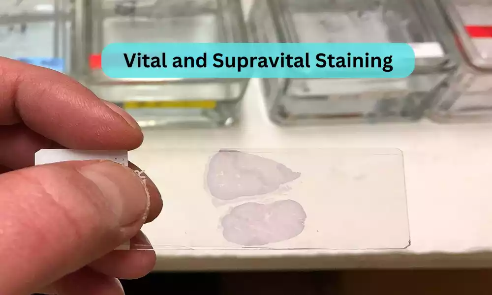
Vital and Supravital Staining are techniques used in microscopy to distinguish living and dead cells, respectively. Vital staining involves applying a dye or stain to living cells or tissues that, under normal conditions, will not kill them. This allows scientists to observe cellular processes in real-time and under natural conditions.
Supravital staining, on the other hand, is applied to cells immediately after removal from an organism, allowing observation of cells that remain functional post-extraction but may not be strictly “alive” in the traditional sense. Both methods offer unique insights into cellular structure and function, providing a window into the dynamic world of cells.
What is Vital Staining?
Vital staining is a technique used in cell biology and histology to study living cells or tissues by using dyes or stains that do not harm or kill them. The term “vital” emphasizes the fact that the process does not significantly interfere with the normal physiology or viability of the cells being studied.
When a vital stain is added to a sample, it allows scientists to observe certain structures, organelles, or processes within living cells. This is particularly useful for monitoring cellular events in real-time or for visualizing specific cell components that might be difficult to distinguish otherwise.
Examples of commonly used vital stains include:
- Trypan blue: Used to determine cell viability. Live cells do not take up the stain, whereas dead cells become blue.
- Neutral red: Taken up by live cells where it accumulates in lysosomes.
- Acridine orange and ethidium bromide: Often used in combination to distinguish between live (green) and dead (red) cells.
- Fluorescein diacetate: Fluoresces in live cells, giving them a greenish appearance.
It’s crucial to select the appropriate vital stain for a given application, as each stain has its unique properties and specificity. Additionally, while vital stains are generally non-toxic at appropriate concentrations, excessive amounts or prolonged exposure can harm cells.
What is Supravital Staining?
Supravital staining is a technique used to study cells immediately after they have been removed from a living organism. Unlike vital staining, which is done on cells or tissues that are still within the living organism or in culture, supravital staining is applied to cells that are no longer in their natural environment but remain metabolically active for a short period after extraction.
The term “supravital” indicates that while the cells are technically outside the living organism and might be in a state of decline, they retain enough functionality to take up the stain. This method allows for the observation of certain cellular processes or structures that might be altered or damaged by more conventional staining techniques that are applied to fixed or dead cells.
A classic example of supravital staining is the:
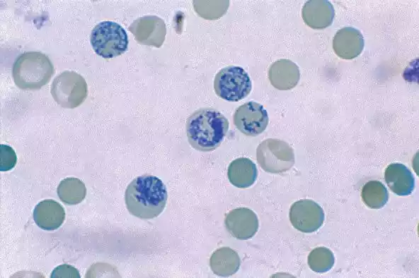
- Reticulocyte count using new methylene blue: Reticulocytes are immature red blood cells. When stained with new methylene blue shortly after blood collection, the reticulum (a network of RNA) becomes visible, aiding in the identification and counting of reticulocytes.
Supravital staining provides a unique perspective on cellular dynamics and structures, bridging the gap between observations in living organisms (via vital staining) and detailed investigations on fixed specimens.
Comparison Table of Vital and Supravital Staining
Here’s a comparison table for Vital and Supravital Staining:
| Aspect | Vital Staining | Supravital Staining |
|---|---|---|
| Definition | Staining of living cells or tissues while they are still within an organism or in culture. | Staining of cells immediately after they have been removed from a living organism. |
| Cell Status | Living cells either in the organism or maintained under life-supporting conditions in culture. | Cells are outside their natural environment but remain metabolically active for a short period. |
| Purpose | To observe cellular processes in real-time within living cells. | To study cells that have just been removed and retain some functionality. |
| Toxicity | Stains are generally non-toxic at the used concentrations. | Cells can tolerate the stain for a short period but might be more susceptible to damage. |
| Applications | – Studying cellular processes in real-time.
– Visualizing specific organelles or structures in living cells. |
– Observing transient cellular events.
– Studying cells that might be altered by conventional staining of dead/fixed cells. |
| Examples | – Trypan blue (for cell viability).
– Neutral red (for lysosomes). |
– New methylene blue (for reticulocyte count). |
This table provides a general overview and comparison between vital and supravital staining. It’s important to note that the specific conditions, techniques, and results can vary based on the exact method and materials used.
How Does Vital and Supravital Staining Work?
Vital and supravital staining are techniques employed to investigate the structure and function of cells, allowing researchers to visualize specific components or processes within the cell. Here’s a brief overview of how these techniques work:
Vital Staining:
- Selectivity: Vital stains are typically selective in what they bind to. For instance, a stain may bind to a specific type of organelle, protein, or other cellular component.
- Permeability: Vital stains must be able to penetrate the cell membrane without causing harm. Once inside the cell, they bind to or get taken up by specific structures.
- Non-toxicity: The chemicals used for vital staining should not be toxic or harmful to the living cells at the concentrations used for staining. This ensures that the natural functions and structures of the cells are not distorted.
- Visualization: Once the stain is inside the cell and has bound to its target, it can be visualized using microscopy. This might involve natural pigmentation of the stain or, in many cases, fluorescence microscopy if fluorescent dyes are used.
Supravital Staining:
- Immediate Application: Supravital stains are applied to cells immediately after their removal from a living organism. Since these cells are still metabolically active, albeit outside their natural environment, they can still take up and process the stains.
- Short-lived Observations: Supravital staining provides a narrow window for observation, given that the cells might begin to deteriorate or lose functionality soon after extraction.
- Selective Binding: Just as in vital staining, supravital stains are often selective, binding to specific cellular components or structures.
- Reduced Toxicity: While the cells in supravital staining are more vulnerable than those in vital staining (given that they’ve been removed from their natural environment), the stains used should still be relatively non-toxic to allow for accurate observations.
- Visualization: Supravital stained cells are observed under a microscope. The observation could involve colorimetric changes, fluorescence, or other visual markers.
The essence of both vital and supravital staining is to gain insights into living cells – whether they are within their natural environment or just removed from it. The choice of technique, stain, and method will largely depend on the specific research question and the type of cells being studied.
Benefits of Vital and Supravital Staining in Biology
Vital and supravital staining have been pivotal tools in biological research, providing scientists with valuable insights into cellular structures, processes, and dynamics in near-natural states. Here are some of the key benefits of these staining techniques in biology:
Benefits of Vital Staining:
- Real-time Observations: Allows researchers to observe cellular processes in real-time, providing insights into the dynamics of living cells.
- Preservation of Cell Function: Since cells remain alive, they continue to carry out their natural functions, enabling studies on metabolism, growth, division, and other processes without the influence of fixation or death.
- Selective Visualization: Many vital stains are selective, binding to specific cellular structures or components, making them prominent for observation.
- Long-term Studies: With appropriate culture conditions, cells can be maintained and observed over extended periods, making it possible to study processes like cell growth, differentiation, and response to stimuli.
- Cell Health Assessment: Some vital stains, like Trypan blue, can differentiate between live and dead cells, helping assess cell viability in cultures.
Benefits of Supravital Staining:
- Transient Cellular Events: Enables the observation of transient events or structures that might be altered or not present in dead or fixed cells.
- Immediate Post-extraction Observations: Provides a snapshot of the cellular state immediately after removal from an organism, capturing processes that might be lost or changed in culture.
- Reduced Artifacts: As the cells are not fixed or subjected to rigorous preparation, artifacts that might distort cellular structures or induce false observations are minimized.
- Detailed Morphological Studies: Especially useful for blood cells, where specific staining can highlight structures (e.g., reticulocyte’s reticular network) that are indicative of cell maturation or function.
- Response to External Factors: Supravital staining can be combined with treatments or stimuli to observe the immediate cellular response, especially if such responses are short-lived.
Both vital and supravital staining techniques bridge the gap between the detailed morphological insights obtained from fixed cells and the dynamic, functional insights obtained from living cells. They allow biologists to study cells in states that are as close to their natural, in vivo conditions as possible, thus providing a more accurate understanding of cellular life.
Limitations and Concerns
While vital and supravital staining offer significant benefits for biological research, they also come with limitations and concerns:
Limitations and Concerns of Vital Staining:
- Potential Toxicity: Even though vital stains are designed to be non-toxic to cells at recommended concentrations, there’s always a possibility that the stain might exert some toxic effects over time or at higher concentrations.
- Interference with Cellular Function: The introduction of a foreign substance (the stain) might influence the normal cellular processes, potentially leading to artifacts.
- Penetration Limitations: Not all stains can efficiently penetrate all cell types or tissues, potentially limiting the scope of observations.
- Non-specific Staining: Some vital stains might bind non-specifically, leading to misleading or ambiguous results.
- Resolution Limitations: Observing living cells, especially in thicker tissues, may offer lower resolution compared to fixed and sectioned tissues, due to light scattering and reduced contrast.
- Maintenance Requirements: Studying living cells requires optimal culture conditions, which might be challenging to maintain over extended periods.
Limitations and Concerns of Supravital Staining:
- Short Observation Window: Cells stained supravitally may begin to deteriorate soon after extraction, providing a limited timeframe for observation.
- Potential Artifacts: Even if cells remain functional immediately after extraction, the act of removing them from their natural environment might induce changes or stresses that can affect observations.
- Difficulty in Handling: Supravital staining often requires rapid and precise handling to ensure cells remain in a suitable state for observation.
- Limited Replicability: Given the transient nature of some observations with supravital staining, certain phenomena might be hard to replicate or might vary between samples.
- Interference from the Dye: The dye itself might interfere with certain cellular processes, especially if it is metabolized or alters the cell’s internal environment.
Both vital and supravital staining techniques demand careful optimization, taking into account the type of cells, the chosen dye, and the specific research question. It’s crucial to be aware of these limitations and plan experiments accordingly, often using complementary methods to confirm and validate observations.
Applications in Modern Research
Vital and supravital staining techniques have seen applications across diverse areas of modern biological and medical research. Their ability to highlight live cellular structures and processes in near-real-time conditions has proven indispensable. Here are some notable applications in modern research:
Applications of Vital Staining:
- Cell Viability Assays: Techniques like the Trypan blue exclusion test help determine the number of viable cells within a cell culture. Such assays are essential in cytotoxicity studies, drug testing, and more.
- Stem Cell Research: Vital staining helps researchers track stem cell differentiation, migration, and integration, especially in live tissue cultures or organoids.
- Neuroscience: Researchers use vital dyes to trace neural pathways, monitor synaptic activity, or visualize specific populations of neurons in live brain slices or in vivo.
- Intracellular Process Tracking: Vital stains can be used to monitor processes like autophagy, endocytosis, or phagocytosis in live cells.
- Fertility and Reproductive Studies: Vital stains are used to assess the viability and health of sperm in fertility clinics.
- Live Cell Imaging: Fluorescent vital dyes, often combined with confocal or multiphoton microscopy, enable researchers to capture dynamic cellular processes in high resolution.
Applications of Supravital Staining:
- Hematology: Supravital stains, like new methylene blue, are employed to visualize reticulocytes in blood samples, helping diagnose and monitor conditions like anemia.
- Transient Cellular Events: Capture and study short-lived cellular events or structures, such as specific metabolic states or immediate responses to external stimuli.
- Cancer Research: Supravital staining can be used to study the immediate effects of radiation or chemotherapy agents on tumor cells extracted from patients.
- Organ Transplant Research: Tissues from donor organs might be subjected to supravital staining to assess cell health and viability before transplantation.
- Drug and Toxicity Studies: Extracted cells can be treated with potential drugs or toxins, followed by supravital staining to rapidly assess effects on cell health or morphology.
These are just a few examples, and the applications are vast and varied. As technologies like microscopy and imaging continue to advance, the potential of vital and supravital staining in modern research is bound to expand further. The techniques provide researchers with powerful tools to visualize and understand the intricacies of life at the cellular level.
Final Thoughts
Vital and supravital staining are critical techniques in cellular biology, facilitating the study of living cells. Vital staining involves introducing a dye or stain to living cells or tissues, illuminating cellular structures without causing immediate harm or cell death. In contrast, supravital staining is applied to cells immediately after their removal from a living organism, offering insights into processes that may be altered post-extraction.
Both methods provide invaluable insights into cellular dynamics, morphology, and function, allowing scientists and researchers to observe biological processes in real-time and contribute to advancements in fields ranging from histology to pathology.